Keyword [PA View & AP View] [Lateral X-ray] [MIMIC-CXR]
Rubin J, Sanghavi D, Zhao C, et al. Large scale automated reading of frontal and lateral chest x-rays using dual convolutional neural networks[J]. arXiv preprint arXiv:1804.07839, 2018.
1. Overview
In this paper, it proposes DualNet
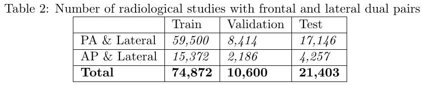
- PA-lateral pair DualNet
- AP-lateral pair DualNet
- experiments on MIMIC-CXR

1.1. Related Work
- ChestX-ray14 dataset
- JSRT dataset
- BSE-JSRT dataset
- Indiana chest X-ray
- Shenzhen dataset
1.2. Limitation
- medical image. 12-bit or greater
- make no distinction between PA and AP
- cardiomegaly can only be accurately assessed in PA image
- AP view will exaggerate the heart silhouette due to magnificention
- lateral view reveals lung areas that are hidden in the frontal view
- lateral view can be useful in detecting lower-lobe lung disease, pleural effusions and anterior mediastinal masses
1.3. Network & Details
- replace 3-channel to 1-channel
- four denseblock (32 growth rate) per layer
- no data augmentation
- Adam with 0.001~0.02 (Triangular2 policy)
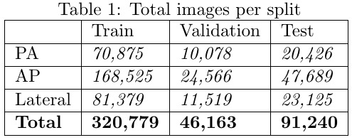
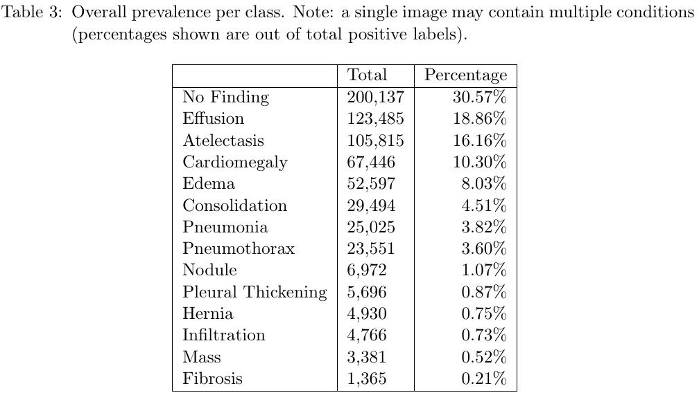
1.4. Dataset
80-20-10
nearest interpolation (ratio mantained). 512x512
- normalize from [0, 2^12-1] to [0, 1]
1.5. Results

- PA results in larger AUC for atelectasis, cardiomegaly, fibrosis, infiltration and pleural thickening
- lateral benefit for consolidation, edema, effusion, hernia, mass, pneumonia and pneumothorax

1.6. Future Work
- improvement. data augmentation, pixel normalization
- patient’s history and current clinical record